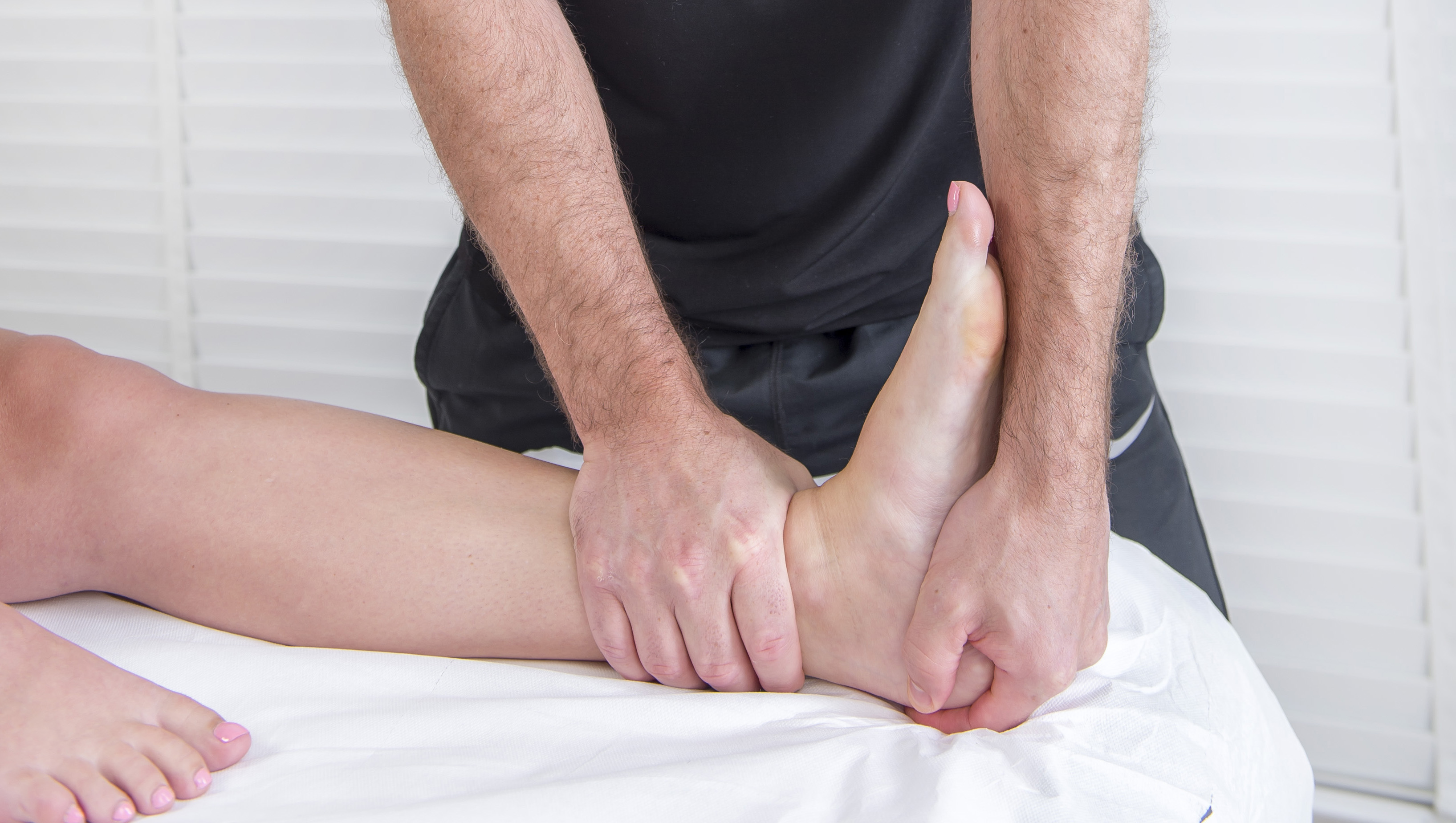
A tibia fracture is a complete or partial break of the shinbone, the large bone that is between ankle and knee. A fracture in this bone is usually caused by trauma, such as a fall or in a car accident. (Repetitive stress can also cause minor cracks in the tibia, occasionally called runner’s stress fracture.) A tibia fracture can be treated in various ways. The best approach for your fracture will depend on various factors, including the severity and type of the break and your normal health. Some fracture needs surgery which is done using orthopedic instruments. We are one of the best trauma implants manufacturers in India.
Nonsurgical Treatment
In some cases, it is possible to treat a tibia fracture without performing surgery. A nonsurgical approach may be considered if there is a slight break and doesn’t extend into a joint. It may also be a suitable option for a patient who has health problems or is less active or who has health that could complicate recovery or surgery. If a nonsurgical treatment is preferable, your doctor may apply a cast to immobilize the bone as well as enable it to heal. After numerous weeks, the cast may be substituted with a brace. You will likely require using crutches to keep weight off the bone as it heals.
Surgery
In most cases, a tibia fracture needs surgery. The goal of tibia fracture surgeries is to rejoin the bone as well as keep it in position as it heals. Most tibia fractures are treated by one of three methods:
- Intramedullary nailing– This is the most common approach for fractures of the tibia. A rod is inserted into the canal that is till the bone center; the rod passes through the fracture to keep the parts in alignment and stable at the time of healing. Screws below and above the fracture secure the rod in position.
- Screws and plates– This approach may be utilized when a fracture extends into the ankle or knee joint. The bones are placed into their correct place; then screws are utilized to attach an orthopedic plate to the outer surface of the bones to hold them in position at the time of healing.
- External fixation– In fractures that involve extensive injury or soft tissue damage, external fixation may be the preferred choice. Screws or metal pins are inserted into the bone near the fracture site through small incisions in the skin. The rod that runs parallel to the bone outside the skin or pins are attached to a bar; this external frame holds the pins in position, so the bone parts cannot move out of place.
The surgery is done using various orthopaedic implants, some are discussed above. There are various orthopedic products manufacturers in India.
What to expect
If you have injured your tibia, your doctor will talk with you about how your injury occurred and will also discover your medical history. After a physical exam, it is likely that you will require one or more imaging test. Possible tests are the following:
- X-ray– An X-ray will give a clear image of the bone and enable the doctor to recognize a break in the tibia, and any type and location of the fracture. An X-ray can also demonstrate if damage has been sustained to joints or neighboring fibula (the smaller long bone in the lower leg).
Computed tomography scan (CT or CAT scan)– A CAT scan may be done if your doctor requires more information. This test uses a computer to combine X-ray images taken from various angles, creating a cross-sectional picture that provides detail. This can be beneficial in visualizing hairline fractures, which may be tough to see on X-ray.


